Biology notes · Chapter 13 of 19
Skeletal & Muscular System
12 sections, 79 flashcards. Open a card to check your answer, or revise them in the app with spaced revision.
Skeleton – Role and Basic Facts
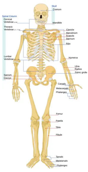
- The skeletal system is the body's internal framework of bones and a few cartilages.
- An adult human has 206 bones plus some cartilages.
- A baby is born with more bones; many fuse during growth to give 206 in the adult.
- Functions: support and shape, protection of soft organs, and movement with muscles.
- Bones store calcium as a mineral reserve.
- The bone marrow inside some bones makes blood cells.
- Osteology is the branch of science that studies bones.
Check yourself
Skeleton – Role and Basic Facts
- The skeletal system is the body's internal framework of bones and a few cartilages.
- An adult human has 206 bones plus some cartilages.
- A baby is born with more bones; many fuse during growth to give 206 in the adult.
- Functions: support and shape, protection of soft organs, and movement with muscles.
- Bones store calcium as a mineral reserve.
- The bone marrow inside some bones makes blood cells.
- Osteology is the branch of science that studies bones.
The is the body's internal framework of bones and a few cartilages.
The skeletal system is the body's internal framework of bones and a few cartilages.
An adult human has plus some cartilages.
An adult human has 206 bones plus some cartilages.
A ; many fuse during growth to give 206 in the adult.
A baby is born with more bones; many fuse during growth to give 206 in the adult.
Functions: , protection of soft organs, and movement with muscles.
Functions: support and shape, protection of soft organs, and movement with muscles.
Bones as a mineral reserve.
Bones store calcium as a mineral reserve.
The inside some bones makes blood cells.
The bone marrow inside some bones makes blood cells.
is the branch of science that studies bones.
Osteology is the branch of science that studies bones.
Connective Tissue – Bone and Cartilage
- Connective tissue binds, supports, strengthens and protects other tissues.
- Its family includes bone, cartilage, blood and fat (adipose).
- Its cells are spread out inside a non-living matrix (ground substance).
- The hardness of the matrix decides how stiff the tissue is.
- In the embryo the skeleton is mostly cartilage, later replaced by bone.
Check yourself
Connective Tissue – Bone and Cartilage
- Connective tissue binds, supports, strengthens and protects other tissues.
- Its family includes bone, cartilage, blood and fat (adipose).
- Its cells are spread out inside a non-living matrix (ground substance).
- The hardness of the matrix decides how stiff the tissue is.
- In the embryo the skeleton is mostly cartilage, later replaced by bone.
binds, supports, strengthens and protects other tissues.
Connective tissue binds, supports, strengthens and protects other tissues.
Its cells are spread out inside a non-living .
Its cells are spread out inside a non-living matrix (ground substance).
The decides how stiff the tissue is.
The hardness of the matrix decides how stiff the tissue is.
In the embryo the skeleton is mostly , later replaced by bone.
In the embryo the skeleton is mostly cartilage, later replaced by bone.
Bone vs Cartilage
- Bone has a hard, non-pliable matrix rich in calcium salts and collagen.
- Cartilage has a soft, pliable matrix with chondroitin salts and little calcium.
- Cartilage can be bent; bone cannot – felt in nose tip and outer ear (pinna).
- Bone cells = osteocytes (in spaces called lacunae); cartilage cells = chondrocytes.
- Cartilage also lies between vertebrae and at the ends of limb bones.
| Feature | Bone | Cartilage |
|---|---|---|
| Matrix | Hard, non-pliable | Soft, pliable |
| Main content | Calcium + collagen | Chondroitin salts |
| Cells | Osteocytes | Chondrocytes |
| Can bend? | No | Yes |
| Found in | Limbs, skull | Nose tip, pinna |
Check yourself
Bone vs Cartilage
- Bone has a hard, non-pliable matrix rich in calcium salts and collagen.
- Cartilage has a soft, pliable matrix with chondroitin salts and little calcium.
- Cartilage can be bent; bone cannot – felt in nose tip and outer ear (pinna).
- Bone cells = osteocytes (in spaces called lacunae); cartilage cells = chondrocytes.
- Cartilage also lies between vertebrae and at the ends of limb bones.
- Matrix — Hard, non-pliable — Soft, pliable
- Main content — Calcium + collagen — Chondroitin salts
- Cells — Osteocytes — Chondrocytes
- Can bend? — No — Yes
- Found in — Limbs, skull — Nose tip, pinna
Bone has a rich in calcium salts and collagen.
Bone has a hard, non-pliable matrix rich in calcium salts and collagen.
Cartilage has a with chondroitin salts and little calcium.
Cartilage has a soft, pliable matrix with chondroitin salts and little calcium.
; bone cannot – felt in nose tip and outer ear (pinna).
Cartilage can be bent; bone cannot – felt in nose tip and outer ear (pinna).
Bone cells = (in spaces called lacunae); cartilage cells = chondrocytes.
Bone cells = osteocytes (in spaces called lacunae); cartilage cells = chondrocytes.
Cartilage also lies and at the ends of limb bones.
Cartilage also lies between vertebrae and at the ends of limb bones.
Two Divisions of the Skeleton
- The skeleton splits by location into axial and appendicular.
- Axial (from axis) = bones of the central midline: head and trunk.
- Appendicular (from appendage) = limb bones plus the two girdles.
- Axial = 80 bones; appendicular = 126 bones.
- Memory check: 80 + 126 = 206 total bones.
| Division | Bones | Examples |
|---|---|---|
| Axial | 80 | Skull, backbone, ribs, sternum |
| Appendicular | 126 | Limbs + pectoral & pelvic girdles |
| Total | 206 | Whole skeleton |
Check yourself
Two Divisions of the Skeleton
- The skeleton splits by location into axial and appendicular.
- Axial (from axis) = bones of the central midline: head and trunk.
- Appendicular (from appendage) = limb bones plus the two girdles.
- Axial = 80 bones; appendicular = 126 bones.
- Memory check: 80 + 126 = 206 total bones.
- Axial — 80 — Skull, backbone, ribs, sternum
- Appendicular — 126 — Limbs + pectoral & pelvic girdles
- Total — 206 — Whole skeleton
The skeleton splits by location into and .
The skeleton splits by location into axial and appendicular.
(from axis) = bones of the central midline: head and trunk.
Axial (from axis) = bones of the central midline: head and trunk.
(from appendage) = limb bones plus the two girdles.
Appendicular (from appendage) = limb bones plus the two girdles.
; appendicular = 126 bones.
Axial = 80 bones; appendicular = 126 bones.
Memory check: total bones.
Memory check: 80 + 126 = 206 total bones.
Skull and Ear Ossicles
- The skull has 22 bones = 8 cranial + 14 facial.
- The 8 cranial bones form the cranium that protects the brain.
- Joints between cranial bones are fixed (immovable) for safety.
- Of the face, only the lower jaw can move; the upper jaw is fixed.
- The U-shaped hyoid bone sits at the base of the mouth.
- Each middle ear has three ossicles – Malleus, Incus, Stapes.
- The stapes is the smallest bone in the body (about 3 mm).
Check yourself
Skull and Ear Ossicles
- The skull has 22 bones = 8 cranial + 14 facial.
- The 8 cranial bones form the cranium that protects the brain.
- Joints between cranial bones are fixed (immovable) for safety.
- Of the face, only the lower jaw can move; the upper jaw is fixed.
- The U-shaped hyoid bone sits at the base of the mouth.
- Each middle ear has three ossicles – Malleus, Incus, Stapes.
- The stapes is the smallest bone in the body (about 3 mm).
The skull has = 8 cranial + 14 facial.
The skull has 22 bones = 8 cranial + 14 facial.
The form the cranium that protects the brain.
The 8 cranial bones form the cranium that protects the brain.
Joints between cranial bones are for safety.
Joints between cranial bones are fixed (immovable) for safety.
Of the face, only the ; the upper jaw is fixed.
Of the face, only the lower jaw can move; the upper jaw is fixed.
The U-shaped bone sits at the base of the mouth.
The U-shaped hyoid bone sits at the base of the mouth.
Each middle ear has three ossicles – .
Each middle ear has three ossicles – Malleus, Incus, Stapes.
Vertebral Column (Backbone)
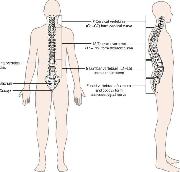
- Built from 26 vertebrae stacked along the back (dorsal) side.
- Each vertebra has a neural canal through which the spinal cord passes.
- The first vertebra, the atlas, joins the skull and lets you nod.
- The atlas–skull joint allows nodding; the axis (pivot joint) allows head rotation.
- Almost all mammals, even the giraffe, have 7 cervical vertebrae.
- The column protects the spinal cord and supports the head.
| Region | Number |
|---|---|
| Cervical (neck) | 7 |
| Thoracic (chest) | 12 |
| Lumbar (lower back) | 5 |
| Sacral (fused) | 1 |
| Coccygeal (fused) | 1 |
| Total | 26 |
Check yourself
Vertebral Column (Backbone)
- Built from 26 vertebrae stacked along the back (dorsal) side.
- Each vertebra has a neural canal through which the spinal cord passes.
- The first vertebra, the atlas, joins the skull and lets you nod.
- The atlas–skull joint allows nodding; the axis (pivot joint) allows head rotation.
- Almost all mammals, even the giraffe, have 7 cervical vertebrae.
- The column protects the spinal cord and supports the head.
- Cervical (neck) — 7
- Thoracic (chest) — 12
- Lumbar (lower back) — 5
- Sacral (fused) — 1
- Coccygeal (fused) — 1
- Total — 26
Built from stacked along the back (dorsal) side.
Built from 26 vertebrae stacked along the back (dorsal) side.
Each vertebra has a through which the spinal cord passes.
Each vertebra has a neural canal through which the spinal cord passes.
The first vertebra, the , joins the skull and lets you nod.
The first vertebra, the atlas, joins the skull and lets you nod.
The atlas–skull joint allows nodding; the allows head rotation.
The atlas–skull joint allows nodding; the axis (pivot joint) allows head rotation.
Almost all mammals, even the giraffe, have .
Almost all mammals, even the giraffe, have 7 cervical vertebrae.
Rib Cage – Ribs and Sternum
- The sternum (breast bone) is a flat bone on the frontmiddle of the chest.
- There are 12 pairs of ribs – thin, flat, curved bones.
- Each rib is bicephalic (two-headed) at its back end.
- True ribs (1–7) join the sternum directly by hyaline cartilage.
- False ribs (8–10) join the 7th rib, not the sternum directly.
- Floating ribs (11–12) are not joined to the sternum in front.
- Thoracic vertebrae + ribs + sternum form the rib cage protecting heart and lungs.
| Rib type | Pairs | Front attachment |
|---|---|---|
| True | 1–7 | Sternum directly |
| False | 8–10 | 7th rib by cartilage |
| Floating | 11–12 | None |
Check yourself
Rib Cage – Ribs and Sternum
- The sternum (breast bone) is a flat bone on the frontmiddle of the chest.
- There are 12 pairs of ribs – thin, flat, curved bones.
- Each rib is bicephalic (two-headed) at its back end.
- True ribs (1–7) join the sternum directly by hyaline cartilage.
- False ribs (8–10) join the 7th rib, not the sternum directly.
- Floating ribs (11–12) are not joined to the sternum in front.
- Thoracic vertebrae + ribs + sternum form the rib cage protecting heart and lungs.
- True — 1–7 — Sternum directly
- False — 8–10 — 7th rib by cartilage
- Floating — 11–12 — None
The is a flat bone on the frontmiddle of the chest.
The sternum (breast bone) is a flat bone on the frontmiddle of the chest.
There are – thin, flat, curved bones.
There are 12 pairs of ribs – thin, flat, curved bones.
join the sternum directly by hyaline cartilage.
True ribs (1–7) join the sternum directly by hyaline cartilage.
join the 7th rib, not the sternum directly.
False ribs (8–10) join the 7th rib, not the sternum directly.
are not joined to the sternum in front.
Floating ribs (11–12) are not joined to the sternum in front.
Thoracic vertebrae + ribs + sternum form the protecting heart and lungs.
Thoracic vertebrae + ribs + sternum form the rib cage protecting heart and lungs.
Appendicular Skeleton – Limbs and Girdles
- A girdle is a ring of bones connecting a limb to the axial skeleton.
- The pectoral girdle attaches the arms: clavicle + scapula.
- The scapula's glenoid cavity holds the humerus to form the shoulder joint.
- The pelvic girdle (hip) attaches the legs; made of two coxal bones.
- Each coxal bone is the fusion of ilium, ischium and pubis.
- Arm bones: humerus, radius, ulna; leg bones: femur, tibia, fibula.
- The femur (thigh bone) is the longest and strongest bone.
| Girdle | Limb | Bones |
|---|---|---|
| Pectoral | Arms | Clavicle + scapula |
| Pelvic | Legs | Two coxal bones |
Check yourself
Appendicular Skeleton – Limbs and Girdles
- A girdle is a ring of bones connecting a limb to the axial skeleton.
- The pectoral girdle attaches the arms: clavicle + scapula.
- The scapula's glenoid cavity holds the humerus to form the shoulder joint.
- The pelvic girdle (hip) attaches the legs; made of two coxal bones.
- Each coxal bone is the fusion of ilium, ischium and pubis.
- Arm bones: humerus, radius, ulna; leg bones: femur, tibia, fibula.
- The femur (thigh bone) is the longest and strongest bone.
- Pectoral — Arms — Clavicle + scapula
- Pelvic — Legs — Two coxal bones
A is a ring of bones connecting a limb to the axial skeleton.
A girdle is a ring of bones connecting a limb to the axial skeleton.
The attaches the arms: clavicle + scapula.
The pectoral girdle attaches the arms: clavicle + scapula.
The scapula's holds the humerus to form the shoulder joint.
The scapula's glenoid cavity holds the humerus to form the shoulder joint.
The attaches the legs; made of two coxal bones.
The pelvic girdle (hip) attaches the legs; made of two coxal bones.
Each is the fusion of ilium, ischium and pubis.
Each coxal bone is the fusion of ilium, ischium and pubis.
Arm bones: ; leg bones: femur, tibia, fibula.
Arm bones: humerus, radius, ulna; leg bones: femur, tibia, fibula.
The is the longest and strongest bone.
The femur (thigh bone) is the longest and strongest bone.
Joints
- A joint is where two or more bones meet.
- Fixed (immovable) joints hold cranial bones together – no movement.
- Slightly movable joints occur between vertebrae.
- Freely movable (synovial) joints allow large movement, e.g. knee, shoulder.
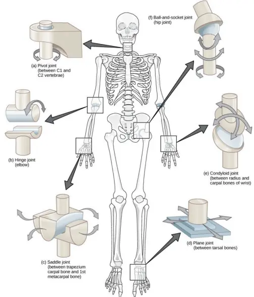
- The ball-and-socket joint (shoulder, hip) moves in all directions.
- The hinge joint (knee, elbow) moves in one plane like a door.
- The pivot joint at the neck (atlas–axis) lets the head turn side to side.
| Joint type | Example | Movement |
|---|---|---|
| Ball-and-socket | Shoulder, hip | All directions |
| Hinge | Knee, elbow | One plane |
| Pivot | Neck (atlas-axis) | Rotation |
| Fixed | Skull | None |
Check yourself
Joints
- A joint is where two or more bones meet.
- Fixed (immovable) joints hold cranial bones together – no movement.
- Slightly movable joints occur between vertebrae.
- Freely movable (synovial) joints allow large movement, e.g. knee, shoulder.
- The ball-and-socket joint (shoulder, hip) moves in all directions.
- The hinge joint (knee, elbow) moves in one plane like a door.
- The pivot joint at the neck (atlas–axis) lets the head turn side to side.
- Ball-and-socket — Shoulder, hip — All directions
- Hinge — Knee, elbow — One plane
- Pivot — Neck (atlas-axis) — Rotation
- Fixed — Skull — None
A is where two or more bones meet.
A joint is where two or more bones meet.
hold cranial bones together – no movement.
Fixed (immovable) joints hold cranial bones together – no movement.
The (shoulder, hip) moves in all directions.
The ball-and-socket joint (shoulder, hip) moves in all directions.
The (knee, elbow) moves in one plane like a door.
The hinge joint (knee, elbow) moves in one plane like a door.
The at the neck (atlas–axis) lets the head turn side to side.
The pivot joint at the neck (atlas–axis) lets the head turn side to side.
The Muscular System
- Muscles are the engines that move the skeleton by contracting.
- Muscle cells are contractile – they can shorten and pull.
- Muscles work in antagonistic pairs (one contracts, the other relaxes).
- Muscles can only pull, not push, so pairs are needed.
- Tendons attach muscle to bone; ligaments join bone to bone.
- The body has over 600 muscles, making up much of body weight.
Check yourself
The Muscular System
- Muscles are the engines that move the skeleton by contracting.
- Muscle cells are contractile – they can shorten and pull.
- Muscles work in antagonistic pairs (one contracts, the other relaxes).
- Muscles can only pull, not push, so pairs are needed.
- Tendons attach muscle to bone; ligaments join bone to bone.
- The body has over 600 muscles, making up much of body weight.
are the engines that move the skeleton by contracting.
Muscles are the engines that move the skeleton by contracting.
Muscle cells are – they can shorten and pull.
Muscle cells are contractile – they can shorten and pull.
Muscles work in (one contracts, the other relaxes).
Muscles work in antagonistic pairs (one contracts, the other relaxes).
Muscles can only , so pairs are needed.
Muscles can only pull, not push, so pairs are needed.
attach muscle to bone; ligaments join bone to bone.
Tendons attach muscle to bone; ligaments join bone to bone.
The body has over , making up much of body weight.
The body has over 600 muscles, making up much of body weight.
Three Types of Muscle
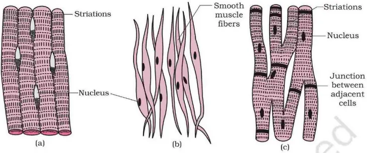
- Skeletal (striated) muscle is voluntary and attached to bones for movement.
- Smooth (unstriated) muscle is involuntary, in walls of stomach and intestines.
- Cardiac muscle is involuntary, found only in the heart, and never tires.
- Skeletal muscle has stripes (striations); smooth muscle does not.
- Cardiac muscle is striated but involuntary – a unique mix.
| Type | Control | Location |
|---|---|---|
| Skeletal | Voluntary | Attached to bones |
| Smooth | Involuntary | Stomach, intestines |
| Cardiac | Involuntary | Heart only |
Check yourself
Three Types of Muscle
- Skeletal (striated) muscle is voluntary and attached to bones for movement.
- Smooth (unstriated) muscle is involuntary, in walls of stomach and intestines.
- Cardiac muscle is involuntary, found only in the heart, and never tires.
- Skeletal muscle has stripes (striations); smooth muscle does not.
- Cardiac muscle is striated but involuntary – a unique mix.
- Skeletal — Voluntary — Attached to bones
- Smooth — Involuntary — Stomach, intestines
- Cardiac — Involuntary — Heart only
is voluntary and attached to bones for movement.
Skeletal (striated) muscle is voluntary and attached to bones for movement.
is involuntary, in walls of stomach and intestines.
Smooth (unstriated) muscle is involuntary, in walls of stomach and intestines.
is involuntary, found only in the heart, and never tires.
Cardiac muscle is involuntary, found only in the heart, and never tires.
Skeletal muscle has ; smooth muscle does not.
Skeletal muscle has stripes (striations); smooth muscle does not.
Cardiac muscle is – a unique mix.
Cardiac muscle is striated but involuntary – a unique mix.
Disorders and Medical Terms
- Osteology is the study of bones.
- Arthritis is inflammation and pain of the joints.
- Osteoporosis is weakening of bones from calcium loss, common in old age.
- Rickets in children is soft, bent bones from vitamin D deficiency.
- Biological resurfacing is a surgical technique to treat cartilage/joint problems.
- A fracture is a break or crack in a bone.
Check yourself
Disorders and Medical Terms
- Osteology is the study of bones.
- Arthritis is inflammation and pain of the joints.
- Osteoporosis is weakening of bones from calcium loss, common in old age.
- Rickets in children is soft, bent bones from vitamin D deficiency.
- Biological resurfacing is a surgical technique to treat cartilage/joint problems.
- A fracture is a break or crack in a bone.
is the study of bones.
Osteology is the study of bones.
is inflammation and pain of the joints.
Arthritis is inflammation and pain of the joints.
is weakening of bones from calcium loss, common in old age.
Osteoporosis is weakening of bones from calcium loss, common in old age.
in children is soft, bent bones from vitamin D deficiency.
Rickets in children is soft, bent bones from vitamin D deficiency.
is a surgical technique to treat cartilage/joint problems.
Biological resurfacing is a surgical technique to treat cartilage/joint problems.
A is a break or crack in a bone.
A fracture is a break or crack in a bone.
All 19 chapters of Biology notes
- Cell: Structure & Function12 sections
- Biomolecules & DNA/RNA11 sections
- Plant Kingdom & Classification11 sections
- Plant Tissues & Anatomy12 sections
- Plant Physiology: Photosynthesis, Respiration & Nutrition14 sections
- Animal Kingdom, Morphology & Classification16 sections
- Nutrition & Digestive System12 sections
- Circulatory System & Body Fluids12 sections
- Respiratory System10 sections
- Excretory System12 sections
- Nervous System & Sense Organs12 sections
- Endocrine System & Hormones14 sections
- Skeletal & Muscular System12 sections
- Reproductive System & Reproductive Health12 sections
- Genetics & Evolution13 sections
- Human Health, Diseases & Immunity16 sections
- Ecology, Ecosystem & Biodiversity12 sections
- Biology in Everyday Life & Applied Biology12 sections
- Inventions, Discoveries & Scientists10 sections