Biology notes · Chapter 8 of 19
Circulatory System & Body Fluids
12 sections, 71 flashcards. Open a card to check your answer, or revise them in the app with spaced revision.
Why We Need a Circulatory System
- The circulatory system is the body's transport network for trillions of cells.
- It has three parts: the heart (pump), blood vessels (pipes) and blood (fluid).
- It delivers oxygen, nutrients, water and hormones to every cell.
- It picks up carbon dioxide (carried to lungs) and wastes (carried to kidneys).
- It defends the body using disease-fighting white blood cells.
- Deep-lying cells cannot exchange directly with surroundings, so a delivery system is essential.
Check yourself
Why We Need a Circulatory System
- The circulatory system is the body's transport network for trillions of cells.
- It has three parts: the heart (pump), blood vessels (pipes) and blood (fluid).
- It delivers oxygen, nutrients, water and hormones to every cell.
- It picks up carbon dioxide (carried to lungs) and wastes (carried to kidneys).
- It defends the body using disease-fighting white blood cells.
- Deep-lying cells cannot exchange directly with surroundings, so a delivery system is essential.
The is the body's transport network for trillions of cells.
The circulatory system is the body's transport network for trillions of cells.
It has three parts: the , and .
It has three parts: the heart (pump), blood vessels (pipes) and blood (fluid).
It picks up (carried to lungs) and wastes (carried to kidneys).
It picks up carbon dioxide (carried to lungs) and wastes (carried to kidneys).
It defends the body using disease-fighting .
It defends the body using disease-fighting white blood cells.
Open vs Closed Circulation
- In an open system, blood leaves vessels and bathes organs directly in spaces called haemocoel.
- The fluid in an open system is haemolymph; flow is slow and pressure is low.
- Open systems are seen in cockroaches and most insects; the cockroach heart has side-holes called ostia.
- In a closed system, blood always stays inside vessels, so flow is fast and pressure stays high.
- All vertebrates and a few invertebrates (like the earthworm) have closed circulation.
- In the earthworm, haemoglobin is dissolved in plasma and its blood cells are phagocytic.
| Feature | Open | Closed |
|---|---|---|
| Blood stays in vessels | No | Yes |
| Fluid name | Haemolymph | Blood |
| Pressure / speed | Low / slow | High / fast |
| Example | Cockroach | Earthworm, vertebrates |
Check yourself
Open vs Closed Circulation
- In an open system, blood leaves vessels and bathes organs directly in spaces called haemocoel.
- The fluid in an open system is haemolymph; flow is slow and pressure is low.
- Open systems are seen in cockroaches and most insects; the cockroach heart has side-holes called ostia.
- In a closed system, blood always stays inside vessels, so flow is fast and pressure stays high.
- All vertebrates and a few invertebrates (like the earthworm) have closed circulation.
- In the earthworm, haemoglobin is dissolved in plasma and its blood cells are phagocytic.
- Blood stays in vessels — No — Yes
- Fluid name — Haemolymph — Blood
- Pressure / speed — Low / slow — High / fast
- Example — Cockroach — Earthworm, vertebrates
In an , blood leaves vessels and bathes organs directly in spaces called .
In an open system, blood leaves vessels and bathes organs directly in spaces called haemocoel.
The fluid in an open system is ; flow is slow and pressure is low.
The fluid in an open system is haemolymph; flow is slow and pressure is low.
Open systems are seen in ; the cockroach heart has side-holes called .
Open systems are seen in cockroaches and most insects; the cockroach heart has side-holes called ostia.
In a , blood always stays inside vessels, so flow is fast and pressure stays high.
In a closed system, blood always stays inside vessels, so flow is fast and pressure stays high.
All and a few invertebrates (like the earthworm) have closed circulation.
All vertebrates and a few invertebrates (like the earthworm) have closed circulation.
In the , haemoglobin is dissolved in plasma and its blood cells are phagocytic.
In the earthworm, haemoglobin is dissolved in plasma and its blood cells are phagocytic.
Heart Chambers Across Animals
- An atrium receives blood returning to the heart; a ventricle pumps blood out.
- Fishes have a 2-chambered heart with single circulation (blood passes the heart once).
- Amphibians and most reptiles have a 3-chambered heart, causing partial mixing of blood.
- The frog heart is wrapped in a pericardium and frogs have a well-developed lymphatic system.
- Crocodiles, birds and mammals have a 4-chambered heart, so blood never mixes.
- The crocodile is the famous reptile exception with 4 chambers.
| Animal | Chambers | Circulation |
|---|---|---|
| Fish | 2 | Single |
| Amphibian / most reptiles | 3 | Incomplete double |
| Crocodile | 4 | Double |
| Bird / mammal | 4 | Double |
Check yourself
Heart Chambers Across Animals
- An atrium receives blood returning to the heart; a ventricle pumps blood out.
- Fishes have a 2-chambered heart with single circulation (blood passes the heart once).
- Amphibians and most reptiles have a 3-chambered heart, causing partial mixing of blood.
- The frog heart is wrapped in a pericardium and frogs have a well-developed lymphatic system.
- Crocodiles, birds and mammals have a 4-chambered heart, so blood never mixes.
- The crocodile is the famous reptile exception with 4 chambers.
- Fish — 2 — Single
- Amphibian / most reptiles — 3 — Incomplete double
- Crocodile — 4 — Double
- Bird / mammal — 4 — Double
An receives blood returning to the heart; a pumps blood out.
An atrium receives blood returning to the heart; a ventricle pumps blood out.
Fishes have a with (blood passes the heart once).
Fishes have a 2-chambered heart with single circulation (blood passes the heart once).
Amphibians and most reptiles have a , causing partial mixing of blood.
Amphibians and most reptiles have a 3-chambered heart, causing partial mixing of blood.
The frog heart is wrapped in a and frogs have a well-developed lymphatic system.
The frog heart is wrapped in a pericardium and frogs have a well-developed lymphatic system.
Crocodiles, birds and mammals have a , so blood never mixes.
Crocodiles, birds and mammals have a 4-chambered heart, so blood never mixes.
The is the famous reptile exception with 4 chambers.
The crocodile is the famous reptile exception with 4 chambers.
Structure of the Human Heart
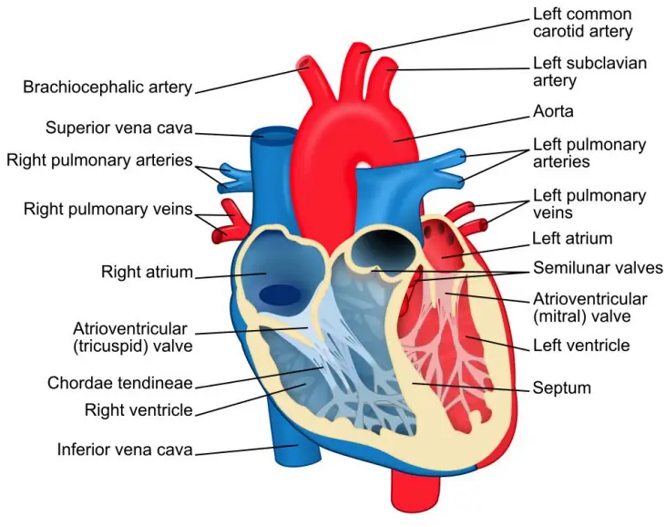
- The human heart is a fist-sized muscular pump lying in the chest, tilted to the left.
- It has four chambers: right atrium, right ventricle, left atrium, left ventricle.
- A muscular wall called the septum keeps oxygen-rich and oxygen-poor blood from mixing.
- The right side handles deoxygenated blood; the left side handles oxygenated blood.
- The left ventricle has the thickest wall to pump blood around the whole body.
- The heart muscle is tireless, contracting and relaxing for life without true rest.
Check yourself
Structure of the Human Heart
- The human heart is a fist-sized muscular pump lying in the chest, tilted to the left.
- It has four chambers: right atrium, right ventricle, left atrium, left ventricle.
- A muscular wall called the septum keeps oxygen-rich and oxygen-poor blood from mixing.
- The right side handles deoxygenated blood; the left side handles oxygenated blood.
- The left ventricle has the thickest wall to pump blood around the whole body.
- The heart muscle is tireless, contracting and relaxing for life without true rest.
The human heart is a lying in the chest, tilted to the left.
The human heart is a fist-sized muscular pump lying in the chest, tilted to the left.
It has : right atrium, right ventricle, left atrium, left ventricle.
It has four chambers: right atrium, right ventricle, left atrium, left ventricle.
A muscular wall called the keeps oxygen-rich and oxygen-poor blood from mixing.
A muscular wall called the septum keeps oxygen-rich and oxygen-poor blood from mixing.
The handles deoxygenated blood; the handles oxygenated blood.
The right side handles deoxygenated blood; the left side handles oxygenated blood.
The heart muscle is , contracting and relaxing for life without true rest.
The heart muscle is tireless, contracting and relaxing for life without true rest.
Heart Valves – One-Way Doors
- Valves are one-way doors that snap shut to stop blood flowing backward.
- The tricuspid valve lies between the right atrium and right ventricle.
- The bicuspid (mitral) valve lies between the left atrium and left ventricle.
- Semilunar valves guard the exits into the pulmonary artery and the aorta.
- The familiar 'lub-dub' heart sounds are the valves snapping shut.
| Valve | Location |
|---|---|
| Tricuspid | Right atrium → right ventricle |
| Bicuspid (mitral) | Left atrium → left ventricle |
| Semilunar | Exits to pulmonary artery & aorta |
Check yourself
Heart Valves – One-Way Doors
- Valves are one-way doors that snap shut to stop blood flowing backward.
- The tricuspid valve lies between the right atrium and right ventricle.
- The bicuspid (mitral) valve lies between the left atrium and left ventricle.
- Semilunar valves guard the exits into the pulmonary artery and the aorta.
- The familiar 'lub-dub' heart sounds are the valves snapping shut.
- Tricuspid — Right atrium → right ventricle
- Bicuspid (mitral) — Left atrium → left ventricle
- Semilunar — Exits to pulmonary artery & aorta
are one-way doors that snap shut to stop blood flowing backward.
Valves are one-way doors that snap shut to stop blood flowing backward.
The lies between the right atrium and right ventricle.
The tricuspid valve lies between the right atrium and right ventricle.
The lies between the left atrium and left ventricle.
The bicuspid (mitral) valve lies between the left atrium and left ventricle.
guard the exits into the pulmonary artery and the aorta.
Semilunar valves guard the exits into the pulmonary artery and the aorta.
The familiar heart sounds are the valves snapping shut.
The familiar 'lub-dub' heart sounds are the valves snapping shut.
Heartbeat, Pacemaker & Cardiac Cycle
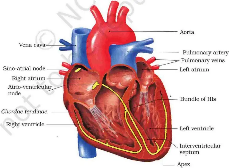
- Heart muscle is auto-excitable – it generates its own electrical signals.
- The sino-atrial node (SAN) in the right atrium is the pacemaker, firing 70–75 times/min.
- Contraction is called systole and relaxation is called diastole.
- One complete heartbeat is a cardiac cycle, lasting about 0.8 seconds.
- An ECG records the heart's electrical activity as P, Q, R, S, T waves.
- A doctor listens to heart sounds with a stethoscope.
Check yourself
Heartbeat, Pacemaker & Cardiac Cycle
- Heart muscle is auto-excitable – it generates its own electrical signals.
- The sino-atrial node (SAN) in the right atrium is the pacemaker, firing 70–75 times/min.
- Contraction is called systole and relaxation is called diastole.
- One complete heartbeat is a cardiac cycle, lasting about 0.8 seconds.
- An ECG records the heart's electrical activity as P, Q, R, S, T waves.
- A doctor listens to heart sounds with a stethoscope.
Heart muscle is – it generates its own electrical signals.
Heart muscle is auto-excitable – it generates its own electrical signals.
The in the right atrium is the , firing 70–75 times/min.
The sino-atrial node (SAN) in the right atrium is the pacemaker, firing 70–75 times/min.
Contraction is called and relaxation is called .
Contraction is called systole and relaxation is called diastole.
One complete heartbeat is a , lasting about .
One complete heartbeat is a cardiac cycle, lasting about 0.8 seconds.
An records the heart's electrical activity as P, Q, R, S, T waves.
An ECG records the heart's electrical activity as P, Q, R, S, T waves.
A doctor listens to heart sounds with a .
A doctor listens to heart sounds with a stethoscope.
Cardiac Output & Key Heart Values
- Stroke volume is the blood pumped per beat by a ventricle, about 70 mL.
- A healthy heart performs about 72 cardiac cycles per minute.
- Cardiac output = stroke volume × heart rate, roughly 5 litres per minute.
- An athlete's cardiac output is higher because stroke volume and heart rate can rise.
- The body increases output during exercise to meet greater oxygen demand.
| Quantity | Normal value |
|---|---|
| Heart rate | ~72 beats/min |
| One cardiac cycle | ~0.8 sec |
| Stroke volume | ~70 mL |
| Cardiac output | ~5 L/min |
Check yourself
Cardiac Output & Key Heart Values
- Stroke volume is the blood pumped per beat by a ventricle, about 70 mL.
- A healthy heart performs about 72 cardiac cycles per minute.
- Cardiac output = stroke volume × heart rate, roughly 5 litres per minute.
- An athlete's cardiac output is higher because stroke volume and heart rate can rise.
- The body increases output during exercise to meet greater oxygen demand.
- Heart rate — ~72 beats/min
- One cardiac cycle — ~0.8 sec
- Stroke volume — ~70 mL
- Cardiac output — ~5 L/min
is the blood pumped per beat by a ventricle, about .
Stroke volume is the blood pumped per beat by a ventricle, about 70 mL.
Cardiac output = stroke volume × heart rate, roughly .
Cardiac output = stroke volume × heart rate, roughly 5 litres per minute.
An is higher because stroke volume and heart rate can rise.
An athlete's cardiac output is higher because stroke volume and heart rate can rise.
Double Circulation – Two Loops
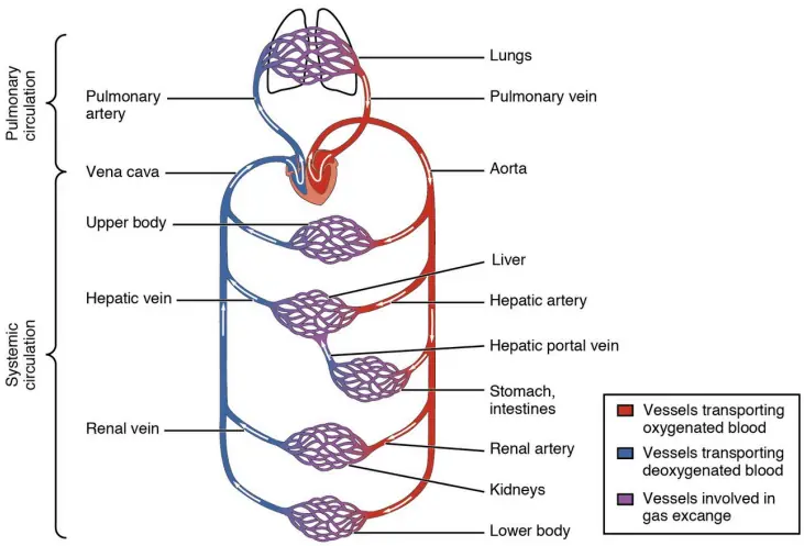
- Double circulation means blood passes through the heart twice per full body trip.
- The pulmonary circuit carries blood from the heart to the lungs and back.
- The systemic circuit carries oxygen-rich blood from the heart to the body and back.
- Mammals and birds have complete double circulation due to a 4-chambered heart.
- This keeps oxygenated and deoxygenated blood fully separated for efficient delivery.
Check yourself
Double Circulation – Two Loops
- Double circulation means blood passes through the heart twice per full body trip.
- The pulmonary circuit carries blood from the heart to the lungs and back.
- The systemic circuit carries oxygen-rich blood from the heart to the body and back.
- Mammals and birds have complete double circulation due to a 4-chambered heart.
- This keeps oxygenated and deoxygenated blood fully separated for efficient delivery.
means blood passes through the heart per full body trip.
Double circulation means blood passes through the heart twice per full body trip.
The carries blood from the heart to the lungs and back.
The pulmonary circuit carries blood from the heart to the lungs and back.
The carries oxygen-rich blood from the heart to the body and back.
The systemic circuit carries oxygen-rich blood from the heart to the body and back.
Blood Vessels – Arteries, Veins, Capillaries
- Arteries carry blood away from the heart and have thick, elastic walls.
- Veins carry blood back to the heart and have valves to prevent backflow.
- Capillaries are the thinnest vessels where exchange of gases and nutrients occurs.
- Most arteries carry oxygenated blood; the pulmonary artery is the exception (carries deoxygenated).
- Most veins carry deoxygenated blood; the pulmonary vein is the only vein carrying oxygenated blood.
- In bypass surgery a vein graft (often the saphenous vein) reroutes blood past blocked arteries.
| Vessel | Blood direction | Exception |
|---|---|---|
| Artery | Away from heart | Pulmonary artery (deoxy.) |
| Vein | Toward heart | Pulmonary vein (oxy.) |
| Capillary | Exchange site | – |
Check yourself
Blood Vessels – Arteries, Veins, Capillaries
- Arteries carry blood away from the heart and have thick, elastic walls.
- Veins carry blood back to the heart and have valves to prevent backflow.
- Capillaries are the thinnest vessels where exchange of gases and nutrients occurs.
- Most arteries carry oxygenated blood; the pulmonary artery is the exception (carries deoxygenated).
- Most veins carry deoxygenated blood; the pulmonary vein is the only vein carrying oxygenated blood.
- In bypass surgery a vein graft (often the saphenous vein) reroutes blood past blocked arteries.
- Artery — Away from heart — Pulmonary artery (deoxy.)
- Vein — Toward heart — Pulmonary vein (oxy.)
- Capillary — Exchange site — –
carry blood away from the heart and have thick, elastic walls.
Arteries carry blood away from the heart and have thick, elastic walls.
carry blood back to the heart and have valves to prevent backflow.
Veins carry blood back to the heart and have valves to prevent backflow.
are the thinnest vessels where exchange of gases and nutrients occurs.
Capillaries are the thinnest vessels where exchange of gases and nutrients occurs.
Most arteries carry oxygenated blood; the is the exception (carries deoxygenated).
Most arteries carry oxygenated blood; the pulmonary artery is the exception (carries deoxygenated).
Most veins carry deoxygenated blood; the is the only vein carrying oxygenated blood.
Most veins carry deoxygenated blood; the pulmonary vein is the only vein carrying oxygenated blood.
Composition of Blood
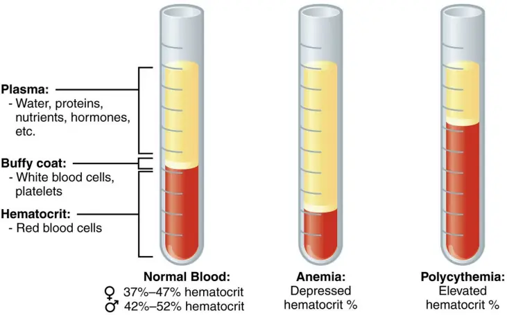
- Blood is a fluid connective tissue made of plasma plus different blood cells.
- Plasma is the liquid part (~55%) carrying water, proteins, salts, nutrients and wastes.
- Red blood cells (RBCs) carry oxygen using the pigment haemoglobin.
- White blood cells (WBCs) fight infection and provide immunity.
- Platelets help blood clot to stop bleeding.
- Total blood volume in a normal adult is about 5 litres.
| Component | Main job |
|---|---|
| Plasma | Liquid transport medium |
| RBC | Carry oxygen (haemoglobin) |
| WBC | Immunity / fight germs |
| Platelet | Blood clotting |
Check yourself
Composition of Blood
- Blood is a fluid connective tissue made of plasma plus different blood cells.
- Plasma is the liquid part (~55%) carrying water, proteins, salts, nutrients and wastes.
- Red blood cells (RBCs) carry oxygen using the pigment haemoglobin.
- White blood cells (WBCs) fight infection and provide immunity.
- Platelets help blood clot to stop bleeding.
- Total blood volume in a normal adult is about 5 litres.
- Plasma — Liquid transport medium
- RBC — Carry oxygen (haemoglobin)
- WBC — Immunity / fight germs
- Platelet — Blood clotting
Blood is a fluid connective tissue made of plus different blood cells.
Blood is a fluid connective tissue made of plasma plus different blood cells.
is the liquid part (~55%) carrying water, proteins, salts, nutrients and wastes.
Plasma is the liquid part (~55%) carrying water, proteins, salts, nutrients and wastes.
carry oxygen using the pigment haemoglobin.
Red blood cells (RBCs) carry oxygen using the pigment haemoglobin.
fight infection and provide immunity.
White blood cells (WBCs) fight infection and provide immunity.
help blood clot to stop bleeding.
Platelets help blood clot to stop bleeding.
Total blood volume in a normal adult is about .
Total blood volume in a normal adult is about 5 litres.
Haemoglobin & Special Blood Facts
- Haemoglobin is an iron-containing red pigment that carries oxygen in blood.
- It gives blood its red colour and does not provide immunity (that is the WBCs' role).
- Some animals (e.g. cockroach, many insects) lack red blood and have colourless 'white' blood.
- Animals like the cockroach have no red blood cells; their haemolymph carries no haemoglobin.
- In the earthworm, haemoglobin is dissolved in plasma rather than packed in cells.
- Low haemoglobin causes anaemia, reducing oxygen delivery.
Check yourself
Haemoglobin & Special Blood Facts
- Haemoglobin is an iron-containing red pigment that carries oxygen in blood.
- It gives blood its red colour and does not provide immunity (that is the WBCs' role).
- Some animals (e.g. cockroach, many insects) lack red blood and have colourless 'white' blood.
- Animals like the cockroach have no red blood cells; their haemolymph carries no haemoglobin.
- In the earthworm, haemoglobin is dissolved in plasma rather than packed in cells.
- Low haemoglobin causes anaemia, reducing oxygen delivery.
is an red pigment that carries oxygen in blood.
Haemoglobin is an iron-containing red pigment that carries oxygen in blood.
It gives blood its red colour and does provide immunity (that is the WBCs' role).
It gives blood its red colour and does not provide immunity (that is the WBCs' role).
Some animals (e.g. cockroach, many insects) lack red blood and have .
Some animals (e.g. cockroach, many insects) lack red blood and have colourless 'white' blood.
Animals like the cockroach have ; their haemolymph carries no haemoglobin.
Animals like the cockroach have no red blood cells; their haemolymph carries no haemoglobin.
In the , haemoglobin is dissolved in plasma rather than packed in cells.
In the earthworm, haemoglobin is dissolved in plasma rather than packed in cells.
Low haemoglobin causes , reducing oxygen delivery.
Low haemoglobin causes anaemia, reducing oxygen delivery.
Blood Groups & Transfusion
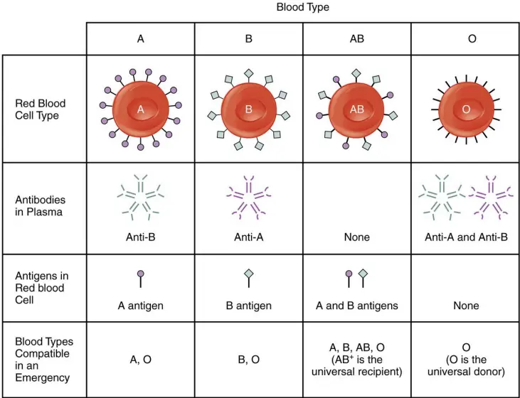
- The ABO system gives four blood groups: A, B, AB and O.
- Group O is the universal donor; group AB is the universal acceptor.
- Blood must match in both ABO type and Rh factor for safe transfusion.
- Rh-positive blood has the Rh antigen; Rh-negative blood lacks it.
- Mismatched transfusion causes clumping (agglutination) of red cells, which can be fatal.
- The Cath lab (catheterization lab) in a hospital is part of the cardiology department.
| Group | Can donate to | Can receive from |
|---|---|---|
| O | All groups (universal donor) | O |
| A | A, AB | A, O |
| B | B, AB | B, O |
| AB | AB | All (universal acceptor) |
Check yourself
Blood Groups & Transfusion
- The ABO system gives four blood groups: A, B, AB and O.
- Group O is the universal donor; group AB is the universal acceptor.
- Blood must match in both ABO type and Rh factor for safe transfusion.
- Rh-positive blood has the Rh antigen; Rh-negative blood lacks it.
- Mismatched transfusion causes clumping (agglutination) of red cells, which can be fatal.
- The Cath lab (catheterization lab) in a hospital is part of the cardiology department.
- O — All groups (universal donor) — O
- A — A, AB — A, O
- B — B, AB — B, O
- AB — AB — All (universal acceptor)
The gives four blood groups: .
The ABO system gives four blood groups: A, B, AB and O.
Blood must match in both for safe transfusion.
Blood must match in both ABO type and Rh factor for safe transfusion.
Rh-positive blood has the ; Rh-negative blood lacks it.
Rh-positive blood has the Rh antigen; Rh-negative blood lacks it.
The (catheterization lab) in a hospital is part of the department.
The Cath lab (catheterization lab) in a hospital is part of the cardiology department.
All 19 chapters of Biology notes
- Cell: Structure & Function12 sections
- Biomolecules & DNA/RNA11 sections
- Plant Kingdom & Classification11 sections
- Plant Tissues & Anatomy12 sections
- Plant Physiology: Photosynthesis, Respiration & Nutrition14 sections
- Animal Kingdom, Morphology & Classification16 sections
- Nutrition & Digestive System12 sections
- Circulatory System & Body Fluids12 sections
- Respiratory System10 sections
- Excretory System12 sections
- Nervous System & Sense Organs12 sections
- Endocrine System & Hormones14 sections
- Skeletal & Muscular System12 sections
- Reproductive System & Reproductive Health12 sections
- Genetics & Evolution13 sections
- Human Health, Diseases & Immunity16 sections
- Ecology, Ecosystem & Biodiversity12 sections
- Biology in Everyday Life & Applied Biology12 sections
- Inventions, Discoveries & Scientists10 sections