Biology notes · Chapter 10 of 19
Excretory System
12 sections, 68 flashcards. Open a card to check your answer, or revise them in the app with spaced revision.
What Is Excretion?
- Excretion is the removal of harmful nitrogen-containing (nitrogenous) metabolic wastes formed inside the body.
- These wastes are leftovers of metabolism (the non-stop chemical work of cells) and would poison the blood if not removed.
- Nitrogenous waste forms when the body breaks down extra proteins and amino acids it cannot store.
- Do not confuse it with egestion – throwing out undigested food as faeces, which was never part of body chemistry.
- Key exam point: only metabolic, nitrogenous waste counts as true excretion.
Check yourself
What Is Excretion?
- Excretion is the removal of harmful nitrogen-containing (nitrogenous) metabolic wastes formed inside the body.
- These wastes are leftovers of metabolism (the non-stop chemical work of cells) and would poison the blood if not removed.
- Nitrogenous waste forms when the body breaks down extra proteins and amino acids it cannot store.
- Do not confuse it with egestion – throwing out undigested food as faeces, which was never part of body chemistry.
- Key exam point: only metabolic, nitrogenous waste counts as true excretion.
is the removal of harmful nitrogen-containing (nitrogenous) metabolic wastes formed inside the body.
Excretion is the removal of harmful nitrogen-containing (nitrogenous) metabolic wastes formed inside the body.
These wastes are leftovers of (the non-stop chemical work of cells) and would poison the blood if not removed.
These wastes are leftovers of metabolism (the non-stop chemical work of cells) and would poison the blood if not removed.
Nitrogenous waste forms when the body breaks down it cannot store.
Nitrogenous waste forms when the body breaks down extra proteins and amino acids it cannot store.
Do not confuse it with – throwing out undigested food as faeces, which was never part of body chemistry.
Do not confuse it with egestion – throwing out undigested food as faeces, which was never part of body chemistry.
Key exam point: only counts as true excretion.
Key exam point: only metabolic, nitrogenous waste counts as true excretion.
Three Types of Nitrogenous Waste
- Ammonia (NH3) is the most toxic but most watersoluble; removed by aquatic animals (ammonotelism).
- Urea is made in the liver from ammonia, is moderately toxic, and is removed by the kidneys.
- Uric acid is least toxic and almost insoluble; thrown out as dry paste to save maximum water (uricotelism).
- Toxicity and water needed both follow the order ammonia > urea > uric acid.
- Humans and frogs are ureotelic (excrete mainly urea).
| Waste | Animal type | Examples |
|---|---|---|
| Ammonia (ammonotelic) | Aquatic | Bony fish, tadpole |
| Urea (ureotelic) | Land mammals | Human, frog |
| Uric acid (uricotelic) | Dry-land | Birds, reptiles, insects |
Check yourself
Three Types of Nitrogenous Waste
- Ammonia (NH3) is the most toxic but most watersoluble; removed by aquatic animals (ammonotelism).
- Urea is made in the liver from ammonia, is moderately toxic, and is removed by the kidneys.
- Uric acid is least toxic and almost insoluble; thrown out as dry paste to save maximum water (uricotelism).
- Toxicity and water needed both follow the order ammonia > urea > uric acid.
- Humans and frogs are ureotelic (excrete mainly urea).
- Ammonia (ammonotelic) — Aquatic — Bony fish, tadpole
- Urea (ureotelic) — Land mammals — Human, frog
- Uric acid (uricotelic) — Dry-land — Birds, reptiles, insects
is the most toxic but most watersoluble; removed by aquatic animals (ammonotelism).
Ammonia (NH3) is the most toxic but most watersoluble; removed by aquatic animals (ammonotelism).
is made in the liver from ammonia, is moderately toxic, and is removed by the kidneys.
Urea is made in the liver from ammonia, is moderately toxic, and is removed by the kidneys.
is least toxic and almost insoluble; thrown out as dry paste to save maximum water (uricotelism).
Uric acid is least toxic and almost insoluble; thrown out as dry paste to save maximum water (uricotelism).
Toxicity and water needed both follow the order .
Toxicity and water needed both follow the order ammonia > urea > uric acid.
Humans and frogs are (excrete mainly urea).
Humans and frogs are ureotelic (excrete mainly urea).
Other Excretory Organs
- Kidneys are the main organs, but they are not the only excretory organs.
- The lungs remove carbon dioxide (CO2) and water vapour.
- The skin removes water, salts and urea through sweat.
- The liver removes bile pigments (bilirubin and biliverdin) from broken-down haemoglobin.
- The large intestine can remove some salts like excess iron and calcium.
| Organ | Waste removed |
|---|---|
| Kidneys | Urea, salts, water (urine) |
| Lungs | CO2, water vapour |
| Skin | Sweat (water, salts, urea) |
| Liver | Bile pigments |
Check yourself
Other Excretory Organs
- Kidneys are the main organs, but they are not the only excretory organs.
- The lungs remove carbon dioxide (CO2) and water vapour.
- The skin removes water, salts and urea through sweat.
- The liver removes bile pigments (bilirubin and biliverdin) from broken-down haemoglobin.
- The large intestine can remove some salts like excess iron and calcium.
- Kidneys — Urea, salts, water (urine)
- Lungs — CO2, water vapour
- Skin — Sweat (water, salts, urea)
- Liver — Bile pigments
Kidneys are the main organs, but they are excretory organs.
Kidneys are the main organs, but they are not the only excretory organs.
The remove carbon dioxide (CO2) and water vapour.
The lungs remove carbon dioxide (CO2) and water vapour.
The removes water, salts and urea through sweat.
The skin removes water, salts and urea through sweat.
The removes bile pigments (bilirubin and biliverdin) from broken-down haemoglobin.
The liver removes bile pigments (bilirubin and biliverdin) from broken-down haemoglobin.
The can remove some salts like excess iron and calcium.
The large intestine can remove some salts like excess iron and calcium.
Parts of the Human Excretory System
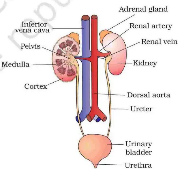
- It has four parts: two kidneys, two ureters, one urinary bladder, one urethra.
- Kidneys are two bean-shaped, reddish-brown organs that filter and clean the blood.
- Ureters are thin tubes carrying urine from each kidney down to the bladder.
- The urinary bladder is a stretchy muscular bag that stores urine.
- The urethra is the tube through which urine finally leaves the body.
Check yourself
Parts of the Human Excretory System
- It has four parts: two kidneys, two ureters, one urinary bladder, one urethra.
- Kidneys are two bean-shaped, reddish-brown organs that filter and clean the blood.
- Ureters are thin tubes carrying urine from each kidney down to the bladder.
- The urinary bladder is a stretchy muscular bag that stores urine.
- The urethra is the tube through which urine finally leaves the body.
are two bean-shaped, reddish-brown organs that filter and clean the blood.
Kidneys are two bean-shaped, reddish-brown organs that filter and clean the blood.
are thin tubes carrying urine from each kidney down to the bladder.
Ureters are thin tubes carrying urine from each kidney down to the bladder.
The is a stretchy muscular bag that stores urine.
The urinary bladder is a stretchy muscular bag that stores urine.
The is the tube through which urine finally leaves the body.
The urethra is the tube through which urine finally leaves the body.
Position and Size of the Kidney
- Each kidney lies between the last thoracic and third lumbar vertebra, against the back wall of the abdomen.
- An adult kidney is about 10-12 cm long, 5-7 cm wide, 2- 3 cm thick.
- Each kidney weighs roughly 120-170 g.
- The inner curved side has a notch, the hilum, where ureter, blood vessels and nerves enter or leave.
- Inside the hilum is a funnel-shaped renal pelvis with finger-like calyces that collect urine.
| Feature | Value |
|---|---|
| Length | 10-12 cm |
| Width | 5-7 cm |
| Thickness | 2-3 cm |
| Weight | 120-170 g |
Check yourself
Position and Size of the Kidney
- Each kidney lies between the last thoracic and third lumbar vertebra, against the back wall of the abdomen.
- An adult kidney is about 10-12 cm long, 5-7 cm wide, 2- 3 cm thick.
- Each kidney weighs roughly 120-170 g.
- The inner curved side has a notch, the hilum, where ureter, blood vessels and nerves enter or leave.
- Inside the hilum is a funnel-shaped renal pelvis with finger-like calyces that collect urine.
- Length — 10-12 cm
- Width — 5-7 cm
- Thickness — 2-3 cm
- Weight — 120-170 g
Each kidney lies between the , against the back wall of the abdomen.
Each kidney lies between the last thoracic and third lumbar vertebra, against the back wall of the abdomen.
Each kidney weighs roughly .
Each kidney weighs roughly 120-170 g.
The inner curved side has a notch, the , where ureter, blood vessels and nerves enter or leave.
The inner curved side has a notch, the hilum, where ureter, blood vessels and nerves enter or leave.
Inside the hilum is a funnel-shaped with finger-like that collect urine.
Inside the hilum is a funnel-shaped renal pelvis with finger-like calyces that collect urine.
Internal Structure of the Kidney
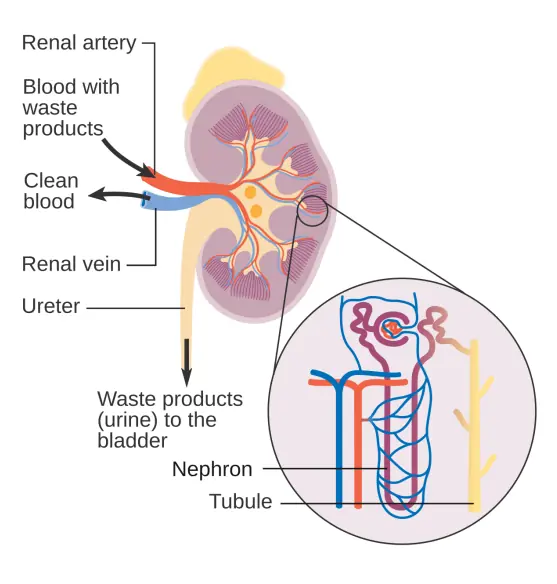
- A tough protective covering, the renal capsule, wraps the kidney from outside.
- A length-wise cut shows two zones: an outer lighter cortex and an inner darker medulla.
- The medulla is divided into cone-shaped medullary pyramids whose tips drip urine into the calyces.
- Cortex tissue dips inward between pyramids as strips called renal columns (columns of Bertini).
- The renal pelvis and calyces collect urine before it enters the ureter.
Check yourself
Internal Structure of the Kidney
- A tough protective covering, the renal capsule, wraps the kidney from outside.
- A length-wise cut shows two zones: an outer lighter cortex and an inner darker medulla.
- The medulla is divided into cone-shaped medullary pyramids whose tips drip urine into the calyces.
- Cortex tissue dips inward between pyramids as strips called renal columns (columns of Bertini).
- The renal pelvis and calyces collect urine before it enters the ureter.
A tough protective covering, the , wraps the kidney from outside.
A tough protective covering, the renal capsule, wraps the kidney from outside.
A length-wise cut shows two zones: an outer lighter and an inner darker .
A length-wise cut shows two zones: an outer lighter cortex and an inner darker medulla.
The medulla is divided into cone-shaped whose tips drip urine into the calyces.
The medulla is divided into cone-shaped medullary pyramids whose tips drip urine into the calyces.
Cortex tissue dips inward between pyramids as strips called .
Cortex tissue dips inward between pyramids as strips called renal columns (columns of Bertini).
The collect urine before it enters the ureter.
The renal pelvis and calyces collect urine before it enters the ureter.
The Nephron - Functional Unit
- Each kidney has over one million nephrons; the nephron is the structural and functional unit of the kidney.
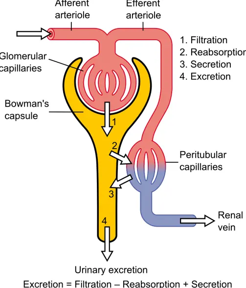
- A nephron has two main parts: the glomerulus (filter) and the renal tubule (winding pipe).
- The glomerulus is a tuft of blood capillaries; blood enters via the afferent and leaves via the efferent arteriole.
- The cup-shaped Bowman's capsule wraps the glomerulus and catches the filtered liquid.
- Glomerulus plus Bowman's capsule form the Malpighian (renal) corpuscle.
- Tubule order: PCT -> Loop of Henle -> DCT -> collecting duct, which empties into the renal pelvis.
Check yourself
The Nephron - Functional Unit
- Each kidney has over one million nephrons; the nephron is the structural and functional unit of the kidney.
- A nephron has two main parts: the glomerulus (filter) and the renal tubule (winding pipe).
- The glomerulus is a tuft of blood capillaries; blood enters via the afferent and leaves via the efferent arteriole.
- The cup-shaped Bowman's capsule wraps the glomerulus and catches the filtered liquid.
- Glomerulus plus Bowman's capsule form the Malpighian (renal) corpuscle.
- Tubule order: PCT -> Loop of Henle -> DCT -> collecting duct, which empties into the renal pelvis.
Each kidney has over ; the nephron is the structural and functional unit of the kidney.
Each kidney has over one million nephrons; the nephron is the structural and functional unit of the kidney.
A nephron has two main parts: the (filter) and the (winding pipe).
A nephron has two main parts: the glomerulus (filter) and the renal tubule (winding pipe).
The is a tuft of blood capillaries; blood enters via the afferent and leaves via the efferent arteriole.
The glomerulus is a tuft of blood capillaries; blood enters via the afferent and leaves via the efferent arteriole.
The cup-shaped wraps the glomerulus and catches the filtered liquid.
The cup-shaped Bowman's capsule wraps the glomerulus and catches the filtered liquid.
Glomerulus plus Bowman's capsule form the .
Glomerulus plus Bowman's capsule form the Malpighian (renal) corpuscle.
Two Types of Nephrons
- Cortical nephrons have a short loop of Henle that barely enters the medulla; they form the majority.
- Juxtamedullary nephrons have a long loop dipping deep into the medulla and make concentrated urine.
- A U-shaped capillary, the vasa recta, runs alongside the long loop and helps save water.
- The vasa recta is poorly developed or absent in cortical nephrons.
| Feature | Cortical | Juxtamedullary |
|---|---|---|
| Loop of Henle | Short | Long |
| Number | Majority | Fewer |
| Vasa recta | Reduced | Well developed |
| Urine concentration | Less | More |
Check yourself
Two Types of Nephrons
- Cortical nephrons have a short loop of Henle that barely enters the medulla; they form the majority.
- Juxtamedullary nephrons have a long loop dipping deep into the medulla and make concentrated urine.
- A U-shaped capillary, the vasa recta, runs alongside the long loop and helps save water.
- The vasa recta is poorly developed or absent in cortical nephrons.
- Loop of Henle — Short — Long
- Number — Majority — Fewer
- Vasa recta — Reduced — Well developed
- Urine concentration — Less — More
have a short loop of Henle that barely enters the medulla; they form the majority.
Cortical nephrons have a short loop of Henle that barely enters the medulla; they form the majority.
have a long loop dipping deep into the medulla and make concentrated urine.
Juxtamedullary nephrons have a long loop dipping deep into the medulla and make concentrated urine.
A U-shaped capillary, the , runs alongside the long loop and helps save water.
A U-shaped capillary, the vasa recta, runs alongside the long loop and helps save water.
The vasa recta is poorly developed or absent in .
The vasa recta is poorly developed or absent in cortical nephrons.
How Urine Is Made - Three Steps
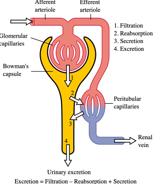
- Step 1 is glomerular filtration: high blood pressure pushes plasma into Bowman's capsule.
- Filtrate passes through endothelium, basement membrane and podocytes with filtration slits, so it is called ultrafiltration.
- Everything except large proteins and blood cells passes through into the capsule.
- Step 2 is reabsorption: useful substances like glucose, amino acids, salts and most water return to blood (mostly in the PCT).
- Step 3 is tubular secretion: extra ions like H+, K+ and ammonia are added into the tubule to keep blood pH balanced.
| Step | What happens |
|---|---|
| Filtration | Plasma filtered into capsule |
| Reabsorption | Useful items returned to blood |
| Secretion | Extra wastes added to filtrate |
Check yourself
How Urine Is Made - Three Steps
- Step 1 is glomerular filtration: high blood pressure pushes plasma into Bowman's capsule.
- Filtrate passes through endothelium, basement membrane and podocytes with filtration slits, so it is called ultrafiltration.
- Everything except large proteins and blood cells passes through into the capsule.
- Step 2 is reabsorption: useful substances like glucose, amino acids, salts and most water return to blood (mostly in the PCT).
- Step 3 is tubular secretion: extra ions like H+, K+ and ammonia are added into the tubule to keep blood pH balanced.
- Filtration — Plasma filtered into capsule
- Reabsorption — Useful items returned to blood
- Secretion — Extra wastes added to filtrate
Step 1 is : high blood pressure pushes plasma into Bowman's capsule.
Step 1 is glomerular filtration: high blood pressure pushes plasma into Bowman's capsule.
Filtrate passes through endothelium, basement membrane and with filtration slits, so it is called .
Filtrate passes through endothelium, basement membrane and podocytes with filtration slits, so it is called ultrafiltration.
Everything except passes through into the capsule.
Everything except large proteins and blood cells passes through into the capsule.
Step 2 is : useful substances like glucose, amino acids, salts and most water return to blood (mostly in the PCT).
Step 2 is reabsorption: useful substances like glucose, amino acids, salts and most water return to blood (mostly in the PCT).
Step 3 is : extra ions like H+, K+ and ammonia are added into the tubule to keep blood pH balanced.
Step 3 is tubular secretion: extra ions like H+, K+ and ammonia are added into the tubule to keep blood pH balanced.
Key Filtration Values (BPSC Numbers)
- About 1100-1200 ml of blood is filtered by the kidneys every minute.
- This is roughly one-fifth (1/5) of the blood the heart pumps each minute.
- GFR (Glomerular Filtration Rate) is about 125 ml of filtrate per minute.
- This adds up to nearly 180 litres of filtrate per day, but only about 1.5 litres becomes urine.
- GFR is regulated by the JGA (Juxtaglomerular Apparatus), which releases the enzyme renin when blood pressure falls.
| Quantity | Value |
|---|---|
| Blood filtered/min | 1100-1200 ml |
| GFR | ~125 ml/min |
| Filtrate/day | ~180 litres |
| Urine/day | ~1.5 litres |
Check yourself
Key Filtration Values (BPSC Numbers)
- About 1100-1200 ml of blood is filtered by the kidneys every minute.
- This is roughly one-fifth (1/5) of the blood the heart pumps each minute.
- GFR (Glomerular Filtration Rate) is about 125 ml of filtrate per minute.
- This adds up to nearly 180 litres of filtrate per day, but only about 1.5 litres becomes urine.
- GFR is regulated by the JGA (Juxtaglomerular Apparatus), which releases the enzyme renin when blood pressure falls.
- Blood filtered/min — 1100-1200 ml
- GFR — ~125 ml/min
- Filtrate/day — ~180 litres
- Urine/day — ~1.5 litres
About is filtered by the kidneys every minute.
About 1100-1200 ml of blood is filtered by the kidneys every minute.
This is roughly of the blood the heart pumps each minute.
This is roughly one-fifth (1/5) of the blood the heart pumps each minute.
This adds up to nearly , but only about 1.5 litres becomes urine.
This adds up to nearly 180 litres of filtrate per day, but only about 1.5 litres becomes urine.
GFR is regulated by the , which releases the enzyme renin when blood pressure falls.
GFR is regulated by the JGA (Juxtaglomerular Apparatus), which releases the enzyme renin when blood pressure falls.
Composition and Passing of Urine
- Normal urine is a light yellow, watery fluid, slightly acidic, with a faint smell.
- It is about 95% water with urea, uric acid, salts, creatinine and the pigment urochrome.
- Glucose, proteins or blood in urine signal disease such as diabetes (glucose) or kidney damage.
- Urine collects in the bladder; its stretching triggers the urge to urinate, called micturition.
- Micturition is controlled by the micturition reflex and relaxation of the urethral sphincters.
Check yourself
Composition and Passing of Urine
- Normal urine is a light yellow, watery fluid, slightly acidic, with a faint smell.
- It is about 95% water with urea, uric acid, salts, creatinine and the pigment urochrome.
- Glucose, proteins or blood in urine signal disease such as diabetes (glucose) or kidney damage.
- Urine collects in the bladder; its stretching triggers the urge to urinate, called micturition.
- Micturition is controlled by the micturition reflex and relaxation of the urethral sphincters.
Normal urine is a , slightly acidic, with a faint smell.
Normal urine is a light yellow, watery fluid, slightly acidic, with a faint smell.
It is about with urea, uric acid, salts, creatinine and the pigment urochrome.
It is about 95% water with urea, uric acid, salts, creatinine and the pigment urochrome.
Glucose, proteins or blood in urine signal disease such as .
Glucose, proteins or blood in urine signal disease such as diabetes (glucose) or kidney damage.
Urine collects in the bladder; its stretching triggers the urge to urinate, called .
Urine collects in the bladder; its stretching triggers the urge to urinate, called micturition.
Micturition is controlled by the and relaxation of the urethral sphincters.
Micturition is controlled by the micturition reflex and relaxation of the urethral sphincters.
Kidney Disorders and Treatment
- Kidney stones (renal calculi) are hard crystals, mainly of calcium oxalate, formed in the kidney.
- Kidney failure means kidneys stop filtering, so urea builds up in blood (uraemia).
- Dialysis is an artificial method to remove wastes from blood when kidneys fail.
- A permanent cure for failure is kidney transplantation from a matching donor.
- Hormones controlling kidney function are ADH (vasopressin), which saves water, and aldosterone, which saves salt.
| Problem | Cause / cure |
|---|---|
| Kidney stone | Calcium oxalate crystals |
| Uraemia | Urea builds in blood |
| Failure | Dialysis or transplant |
Check yourself
Kidney Disorders and Treatment
- Kidney stones (renal calculi) are hard crystals, mainly of calcium oxalate, formed in the kidney.
- Kidney failure means kidneys stop filtering, so urea builds up in blood (uraemia).
- Dialysis is an artificial method to remove wastes from blood when kidneys fail.
- A permanent cure for failure is kidney transplantation from a matching donor.
- Hormones controlling kidney function are ADH (vasopressin), which saves water, and aldosterone, which saves salt.
- Kidney stone — Calcium oxalate crystals
- Uraemia — Urea builds in blood
- Failure — Dialysis or transplant
are hard crystals, mainly of calcium oxalate, formed in the kidney.
Kidney stones (renal calculi) are hard crystals, mainly of calcium oxalate, formed in the kidney.
means kidneys stop filtering, so urea builds up in blood (uraemia).
Kidney failure means kidneys stop filtering, so urea builds up in blood (uraemia).
is an artificial method to remove wastes from blood when kidneys fail.
Dialysis is an artificial method to remove wastes from blood when kidneys fail.
A permanent cure for failure is from a matching donor.
A permanent cure for failure is kidney transplantation from a matching donor.
Hormones controlling kidney function are , which saves water, and aldosterone, which saves salt.
Hormones controlling kidney function are ADH (vasopressin), which saves water, and aldosterone, which saves salt.
All 19 chapters of Biology notes
- Cell: Structure & Function12 sections
- Biomolecules & DNA/RNA11 sections
- Plant Kingdom & Classification11 sections
- Plant Tissues & Anatomy12 sections
- Plant Physiology: Photosynthesis, Respiration & Nutrition14 sections
- Animal Kingdom, Morphology & Classification16 sections
- Nutrition & Digestive System12 sections
- Circulatory System & Body Fluids12 sections
- Respiratory System10 sections
- Excretory System12 sections
- Nervous System & Sense Organs12 sections
- Endocrine System & Hormones14 sections
- Skeletal & Muscular System12 sections
- Reproductive System & Reproductive Health12 sections
- Genetics & Evolution13 sections
- Human Health, Diseases & Immunity16 sections
- Ecology, Ecosystem & Biodiversity12 sections
- Biology in Everyday Life & Applied Biology12 sections
- Inventions, Discoveries & Scientists10 sections